


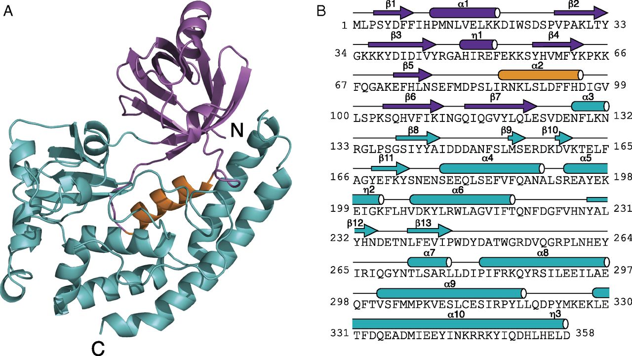
Fig. 3.
Crystal structure of B. cereus CotH reveals an atypical PKL fold. (A) Ribbon representation of B. cereus CotH. The N- and C-lobes are shown in magenta and teal, respectively. The α2 helix (PKA αC equivalent) is highlighted in orange. (B) Amino acid sequence of B. cereus CotH depicting the secondary structural elements, color-coded as in A. The 310 helices (η) are also shown.

