


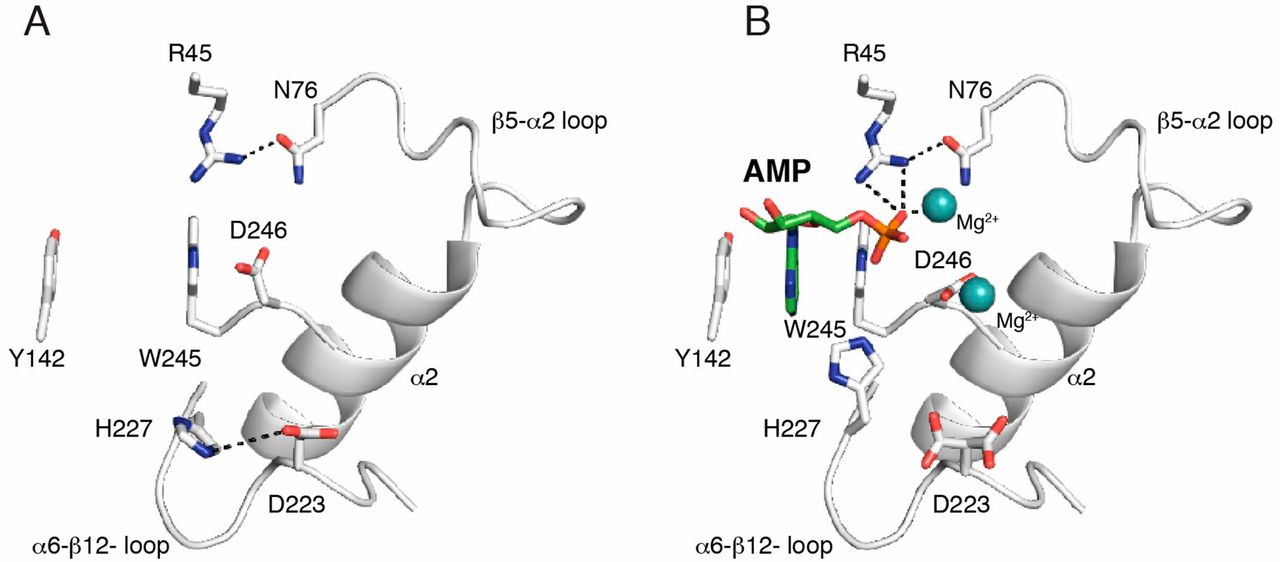
Fig. S4.
Comparison of the active sites in the apo- and AMP/Mg2+ structures. Expanded images of the nucleotide-binding pocket in the apo structure (A) and the AMP/Mg2+ structure (B) are shown. The AMP molecule and the two Mg2+ ions are shown as green sticks and cyan spheres, respectively. Interactions are shown as dashed lines.

