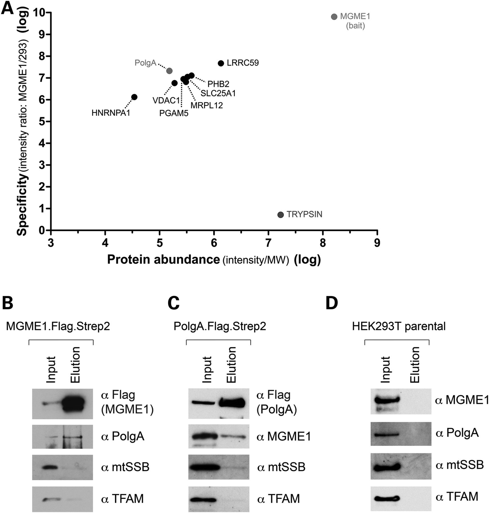
Interaction of MGME1 and PolgA. (A) Mitochondria from parental HEK293T cells or derivatives expressing MGME1.Flag were isolated, lysed, and protein extracts were subjected to affinity purification using anti-Flag beads. Graph presents quantitative analysis of MS data obtained on eluates. Protein names are displayed. MGME1, PolgA and trypsin are highlighted. The entire dataset is specified in Supplementary Material, Table S5. (B) Western blot result of pull-down of the endogenous PolgA with MGME1.Flag.Strep2. The mitochondrial lysates (‘Input’) and eluates (‘Elution’) were analysed with the antibodies as indicated. (C) Reciprocal western blot confirmation of the MGME1–PolgA interaction by co-IP of the endogenous MGME1 with PolgA.Flag.Strep2. (D) Western blot of the control experiment performed using parental HEK293T cells, which do not express either recombinant MGME1 or PolgA proteins.


